PARTIAL PENECTOMY IN THE FIELD IN EQUINE FOR SQUAMOUS CELL CARCINOMA: CASE REPORT
REGISTRO DOI: 10.69849/revistaft/ra10202502162039
Daniely Da Silva Barbosa1
Francisco Alessandro De Araújo Rodrigues Júnior2
Jucimara Santana Carneiro3
Jovanna Karine Pinheiro4
Helizadora Magalhães Rosado Monteiro5
Luana Vieira Cruz6
Anne Gabrielle Moura Ferreira7
Isadora Gomes Noca Bezerra Almeida8
Annielle Regina Da Fonseca Fernandes9
Vinicius Tenório Máximo10
Resumo – O carcinoma espinocelular é uma neoplasia maligna de células escamosas que afeta os queratinócitos, com alto índice de metástase e de importância significativa na rotina clínica e cirurgia de equinos, denominado também de carcinoma de células escamosas, possui predileção por animais idosos e não castrados. O presente relato baseia-se no caso clínico de um equino; macho; 14 anos de idade; pesando 250 kg; resgatado e atendido na cidade de Juazeiro do Norte – CE. Como queixa principal foi relatado a presença de uma massa neoplásica na extremidade distal do pênis com presença de focos difusos de ulceração. O diagnóstico foi baseado nos exames clínico e histopatológico com resultados compatíveis com carcinoma de células escamosas. O animal foi submetido ao tratamento cirúrgico no qual foi utilizado a técnica de penectomia parcial realizada à campo com uso de anestesia infiltrativa. A exérese foi realizada com sucesso, sem intercorrências no trans e pós cirúrgico, o animal teve alta e a recuperação ocorreu sem mais complicações decorrentes do procedimento.
Palavras-chave: Carcinoma. Neoplasia. Histopatológico.
Abstract – Squamous cell carcinoma is a malignant neoplasm affecting squamous cells, with a high metastatic rate and significant importance in equine clinical and surgical routines. Also known as squamous cell carcinoma, it primarily affects older, non-castrated animals. This report is based on the clinical case of a 14-year-old, 250 kg male horse rescued and treated in Juazeiro do Norte – CE. The main complaint was the presence of a neoplastic mass at the distal end of the penis with diffuse ulceration. Diagnosis was based on clinical and histopathological examinations, which showed results consistent with squamous cell carcinoma. The animal underwent surgical treatment using the partial penectomy technique performed in the field under infiltrative anesthesia. The excision was successfully performed without intraoperative or postoperative complications, and the animal was discharged with recovery occurring without further procedure-related issues.
Keywords: Carcinoma. Neoplasia. Histopathological.
Squamous cell carcinoma is a malignant and metastatic skin neoplasm of keratinocytes (Scattaregi, 2017), which can be found in various areas of the body, with a predisposition for regions with sparse hair and lower skin pigmentation (Rabbers et al., 2014). Affected animals may present with increased volume in the penile and preputial regions, urinary retention, and urethral canal involvement (Xavier, 2008). Surgical treatment involves tumor resection through penectomy. The aim of this study is to report a case of squamous cell carcinoma, as well as the surgical technique and anesthetic protocol used for partial penectomy performed in the field on an equine.
A 14-year-old, castrated, mixed-breed male horse, with a body condition score of II, weighing 250 kg, with chestnut coat color, was rescued in Juazeiro do Norte-CE and referred to a regional hospital on 11/10/2023 with a tumor in the penile region, where it was initially treated only with clinical management. On physical examination, bilateral mucopurulent nasal discharge was noted without pulmonary changes. In the pubic region, a mass was observed at the distal end of the penis with the presence of serosanguineous secretion and a foul odor (Figures 1 and 2).
The complete blood count revealed mild anemia. A biopsy sample of the tumor was collected and immersed in 10% formalin by excisional method, showing multilobulated neoplastic characteristics, surrounded by hairy skin, with diffuse foci of ulceration, measuring 12 x 14 cm, for histopathological examination. Microscopy revealed a nonencapsulated, infiltrative neoplasm composed of cords or islands of squamous epithelial cells in the dermal and subcutaneous regions. The neoplastic cells had large, ovoid, often vesicular nuclei containing one or more prominent basophilic nucleoli, abundant cytoplasm varying from amphophilic to eosinophilic, and distinct cell borders. There were keratin pearls, marked anisocytosis and anisokaryosis, with eight mitotic figures in 10 consecutive fields at 400x magnification. Ulceration was noted in the epidermal region with associated inflammatory presence linked to a bacterial infectious process (cocci). The histopathological findings were consistent with a diagnosis of well-differentiated squamous cell carcinoma.
Medical treatment was provided for secondary infection in the nasal region with dexamethasone (12.5ml; IV; ⅓), flunixin (5ml; IV; ⅓), and enrofloxacin (12.5ml; IM; 1/7). Subsequently, the patient was referred for partial penectomy in the field. During the anesthetic procedure, acepromazine (0.02mg/kg) and detomidine (0.02mg/kg) were administered as pre-anesthetic medication (PAM). Induction was performed with midazolam (0.15mg/kg) and ketamine (1.5mg/kg). Lidocaine (0.1ml/kg per point) was used for local pudendal nerve block, and maintenance was achieved via intravenous triple-drip: xylazine (0.5mg/ml), ketamine (2mg/ml), and GGE (50mg/ml) in continuous infusions of 2ml/kg/h.
The animal was restrained in right lateral recumbency. The surgical procedure began with urethral catheterization to relieve the obstructed urine flow (Figure 3). To prevent hemorrhage, a tourniquet was applied proximally at the base of the penis. A triangular incision was made in the tunica albuginea and the corpus cavernosum of the penis up to the urethra, where the urethral catheter could be visualized. After excision of the neoplastic mass and hemostasis of the blood vessels, the edges of the urethra and penile fibrous tissue were sutured using 2-0 Vicryl in a simple interrupted pattern (Figure 4). Subsequently, the cavernous and spongy structures at the end of the penis were sutured in a simple interrupted pattern with 2-0 Vicryl, and the tourniquet was then removed, leaving only the urethral catheter in place (Figure 5).
Squamous cell carcinoma (SCC) is a cutaneous and invasive neoplasm that has a predisposition for lightly pigmented areas and often affects castrated males, as the accumulation of smegma produced by the preputial gland can be exacerbated by solar radiation (Xavier, 2010). The combination of clinical signs may include skin thickening, mild exfoliation followed by epidermal thinning, and ulceration (Rocha, 2010). Erosive lesions that destroy the overlying epidermis can lead to ulceration, necrosis, and a strong odor (Pascoe & Knottenbelt, 1999). Although macroscopic characteristics of the lesion and the animal’s age provide important clues (Wright & Delaunois-Vanderperren, 2010), the definitive diagnosis of neoplasms in animals generally relies on histopathological examination (Wright & Delaunois-Vanderperren, 2010). Differential diagnoses may include sarcoid, pythiosis, habronemiasis, exuberant granulation tissue, stephanofilariasis, papilloma, and fibropapilloma (McGavin and Zachary, 2009; Coelho et al., 2012).
Penectomy is the only effective surgical method in cases of neoplasia (Ferreira, 2019). According to Turner (2002), recommendations for total or partial procedures are associated with the extension of the penis volume with no prognosis of reversal. In this case, the technique was indicated due to the involvement of the urethral canal and the risk of metastasis, and it was decided to perform the surgery in the field using infiltration anesthesia and maintenance with triple-drip. It has been reported that horses anesthetized with triple-drip exhibit fewer negative effects related to cardiorespiratory function and better tissue perfusion conditions due to maintaining adequate blood pressure, providing better analgesia, and loss of reflexes (Hellu, 2012; Beths, 2007).
This case highlights the importance of vigilance and early intervention in managing SCC in horses. The decision for penectomy surgery underscored the need for aggressive therapeutic approaches to control neoplasia and reduce the risk of complications and metastasis. The surgical technique and anesthesia protocol used proved effective, as tumor excision was successfully performed without intraoperative complications. There are no records in the literature of other methods for performing penectomy. Finally, the animal was released and returned to the care of its owner.
1. References
BATAIER, M. N. et al. Carcinoma de células escamosas em prepúcio de equino: relato de caso. Revista Científica Eletrônica de Medicina Veterinária, Garça, v. 9, n. 18, 2012. Disponível em: https://bit.ly/2PIttR8. Acesso em: 18 fev. 2018.
BETHS, T. Total intravenous techniques for anaesthesia. In Practice, v. 29, p. 410413, 2007.
COELHO, C.M.M.; SILVA, O.C.; SILVA, L.A. F.; RABELO, R.E.; ORLANDO, C.F.P.; ARAÚJO, I.L.F. Enfermidades cirúrgicas do aparelho reprodutor masculino equino: aspectos clínicos e terapêuticos. Revista do Conselho Federal de Medicina Veterinária, v.18, n. 55, p. 58-66, 2012.
FERREIRA, D. T. Penectomia parcial em equino: relato de caso. 2019. Trabalho de
Conclusão de Curso (Bacharelado em Medicina Veterinária) – Centro Universitário Luterano de Palmas, Palmas, Tocantins, 2019. Disponível em: http://ulbrato.br/bibliotecadigital/publico/home/documento/1404. Acesso em: 17 abr. 2024.
HELLU, J. A. A.; NETO, I. M.; DUQUE, J. C. M. Avaliação da segurança e eficácia clínica de uma solução líquida de éter gliceril guaiacol pronta para uso (egg-ppu) em cavalos. Ars Veterinaria, v. 28, n. 4, p. 209-217, 2012.
OPORTO, C. I. S. et al. Penectomia parcial em equino com carcinoma espinocelular: relato de caso. Revista de Educação Continuada em Medicina Veterinária e Zootecnia do CRMV-SP, v. 16, n. 3, p. 60-68, 11 dez. 2018.
RABBERS, A. S., et al. Diagnóstico clínico, laboratorial e tratamento cirúrgico do carcinoma de células escamosas no genital de equinos machos: relatos de dois casos. Revista Brasileira de Ciência Veterinária, 21(1):12-18, 2014.
ROCHA, J. R.; SANTOS, L. M.; TRENTIN, T. C.; ROCHA, F. P. C.; PACHECO, M. D. Carcinoma de Células Escamosas em Cães – Relato de Caso. Revista Científica Eletrônica de Medicina Veterinária, n. 14, 2010.
SCATTAREGI, C. S. et al. Penectomia parcial em equinos com carcinoma espinocelular. Revista Acadêmica de Ciência Animal, v. 15, p. 333-333, 2017.
SCOTT, D.W., MILLER, W. H. J.; Dermatologia Eqüina. Intermedica Editorial XXI2004. Buenos Aires – República Argentina, p. 625, 2004.
TURNER, A. S.; MCILWRAITH, C. W. Técnicas cirúrgicas em animais de grande porte. Fort Collins, Colorado: Roca, 2002. 331 p.
WRIGHT, B.; DELAUNOIS-VANDERPERREN, H. Tumours and Tumour-like Growths in Horses – Neoplastic Masses, 2010.
XAVIER, F. S. et al. Estudo retrospectivo e preliminar de carcinomas de células escamosas em trato genital masculino em equinos, durante o período de 1983 a 2008. In: X Encontro de Pós-Graduação FV/UFPel. 2008.
Xavier, F. S. (2010). Lesões proliferativas de pênis e prepúcio eqüinos. Universidade Federal de Pelotas, Pelotas, Rio Grande do Sul.
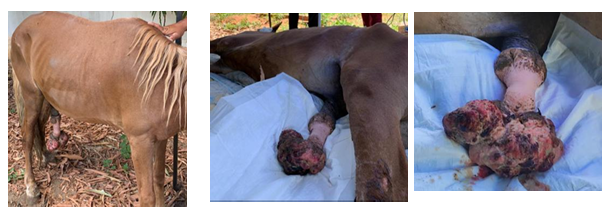
Figure 1– Animal standing, presenting a tumor at the distal end of the penis. Photo: Jucimara Santana Carneiro.
Figure 2 A and B– Animal sedated and in right lateral recumbency; tumor at the distal end of the penis presenting a serosanguineous discharge. Photo: Daniely da Silva Barbosa.
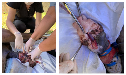
Figure 3– Urethral catheterization being performed to clear the urethral canal and restore urine flow. Photo: Daniely da Silva Barbosa.
Figure 4 – Suturing of the urethral edges and fibrous tissue using 2-0 Vicryl in a simple interrupted pattern after neoplasm excision. Photo: Daniely da Silva Barbosa.
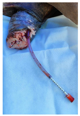
Figure 5– Suturing of the cavernous and spongy structures at the distal end of the penis using 2-0 Vicryl in a simple interrupted pattern, with a urethral catheter in place. Photo: Vinicius Tenório Máximo.
1Discente do Centro Universitário Maurício de Nassau – UNINASSAU – Juazeiro do
Norte – CE. Email: dsbarbosa.aluno@gmail.com
2Discente do Centro Universitário Maurício de Nassau – UNINASSAU – Juazeiro do Norte -CE
Email: Junior.crato201@gmail.com
3Discente do Centro Universitário Maurício de Nassau – UNINASSAU – Juazeiro do Norte – CE
Email: marasantanaballet1@gmail.com
4Docente do Centro Universitário Vale do Salgado – UNIVS – Icó – CE
Email: jovannakarine@univs.edu.br
5Discente do Centro Universitário Maurício de Nassau – UNINASSAU – Juazeiro do Norte – CE
Email: 37015440@sempreunijuazeiro.com.br
6Discente do Centro Universitário Maurício de Nassau – UNINASSAU – Juazeiro do Norte – CE
Email: 370101035@prof.unijuazeiro.edu.br
7Médica Veterinária autônoma
Email: 201910069@acad.unijuazeiro.edu.br
8Médica Veterinária responsável técnica da clínica escola de medicina veterinária –
UNINASSAU – Juazeiro do Norte – CE
Email: Isadoragnba@gmail.com
9Docente da Unidade Acadêmica de Medicina Veterinária/Universidade Federal de
Campina Grande (UAMV/UFCG)
anni.regina@gmail.com
10Docente especialista do Centro Universitário Maurício de Nassau – UNINASSAU – Juazeiro do Norte – CE
Email: 370100945@profunijuazeiro.edu.br