REGISTRO DOI: 10.5281/zenodo.8132618
Jennifer Hummel1
Gustavo Vicente1
Isamery A Machado de Sarmiento2,3
Maria J Pereira de Campo4
Guilherme L Savassi Rocha5
Fernando Carmona Dinau6
Fabiane Zanchin1
Noeme Sousa Rocha6
ABSTRACT
Multiple exostosis (ME) is a disease that can affect the musculoskeletal system of different species. In canines, it presents benignly and infrequently, but also its etiology is still poorly understood. ME has been described in dogs after the growth phase as being related to an alteration of the EXT1 or EXT2 genes, associated with the secretion of heparan sulfate, a connective tissue component necessary for developing cartilage and bone. We report a case of multiple exostosis in a six-month-old Akita dog. Some clinical signs such as thoracolumbar pain, ataxia, and paraparesis, were detected. The patient manifested an increased patellar reflex in the lower limb and bilateral patellar dislocation during the examination. Thoracolumbar tomography revealed a thickening of the L1 vertebra, indicating an aberrant bone formation in the spinous process in its distal region, which caused spinal cord and nerve root compression. Decompressive corpectomy surgery removed abnormal bone growths, improving the symptoms.
Keywords: canine, biopsy, EXT1, and EXT2 genes, multiple exostosis, tomography.
Multiple exostosis (ME) is a benign disease that affects the locomotor system. ME has also been called osteochondromatosis, osteocartilaginous exostosis, osteochondroma, hereditary dyschondroplasia deformans, and diaphyseal aclasis,9 characterized by a proliferative condition in bone and cartilage. Abnormal cartilage growth with bone overlay can be noted in the epiphyseal or subarticular plates.4 This proliferative condition, where the type of endochondral ossification is located, can occur in vertebrae, rib, long bones, and pelvis of animals like dogs, horses, and cats, being more frequent in humans. In some cases, this benign disease can transform into malignant.13 Literature poorly describes ME in canine, only in cases after the growth phase. Here, it is essential to highlight that connective tissue components, specifically proteoglycan, have been associated with a heparan sulfate (HS) problem. HS is responsible for regulating the development of cartilage and bone by interacting with bone proteins.1 Such specific mechanisms of this disease are still unknown. However, some authors have recently described a mutation in the EXT1 or EXT2 genes that encode the glycosyltransferase enzyme responsible for the synthesis of HS.3,7,11,12
The treatment of choice for exostosis, especially in cases where there is a nerve compression or pain, is surgery. However, a study conducted in rats showed that Palovarotene, a retinoic acid receptor selective agonist, can inhibit the formation of osteochondroma in rats with the EXT 1 gene mutation.14 Other therapeutic targets are also being studied, such as heparanase, the enzyme responsible for breaking the HS chain. Inhibitors of this enzyme such as SST0001 have shown promising in vitro results in preventing disease progression in humans.10
A 6-month-old male canine, Akita breed, 20 kg of weight, was admitted for consultation. The clinical examination revealed paraparesis, ataxia, and thoracolumbar pain. After three months, it presented paraplegia. Clinical signs at the consultation time were increased patellar reflex in pelvic limbs and bilateral patellar dislocation. The laboratory tests regarding hematology and blood chemistry were within normal ranges, with a slight platelet decrease. Subsequently, radiology examinations were performed on the right and left pelvic limbs, medially and laterally, and in the abdominal pelvic region, ventrodorsal position. Concerning the left coxofemoral joint, a slight thickening was observed in the neck of the femur, with signs of dislocation.
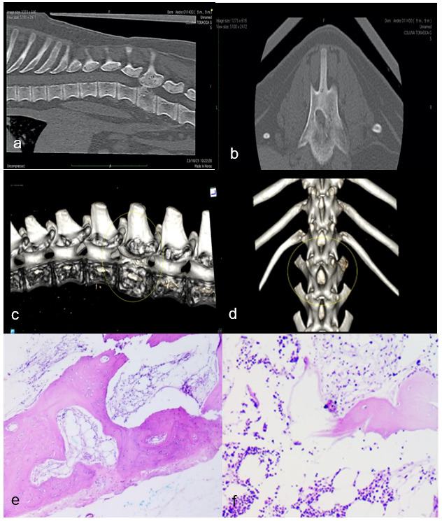
In the tomography examination of the thoracolumbar region, in different views (Fig. 1a and 1b), thickening of the L1 vertebra was observed, which shows abnormal bone growth in the region of the spinous process and distal region of the vertebra, which caused compression of the spinal cord and nerve root.
Figure 1

Finally, a decompressive corpectomy surgery was performed on the affected vertebra, and biopsy samples were taken and sent for histopathological study. Differentiated bone tissue was observed with lacunae and osteocytes, hyaline cartilage with endochondral-type bone growth zones, and hematopoietic tissue (Fig. 1e and 1f).
After performing all the exams, the Multiple Exostosis diagnosis coincided with the characteristics reported by different authors. This disease has also been called osteochondromatosis, osteocartilaginous exostosis, osteochondroma, hereditary dyschondroplasia deformans, and diaphyseal aclasis.2,4
Multiple exostosis has been characterized by irregular bone formation at the level of the epiphyseal or subarticular plates that infrequently occurs in various animals such as horses, cats, dogs, and frequently in humans.13 The age for its appearance is in the development stage, the phase where the musculoskeletal system is fully developed.4,6,14 It coincides with the dog evaluated in this case report, aged six months old.
The symptomatology presented by the patient in this case report was thoracolumbar pain, ataxia, paraparesis, and, after three months, paraplegia. Different exams were performed, including radiographs, ultrasound scans, and, later in time, spinal decompression surgery. As for the complete hematology and blood chemistry, the exams were in the normal range, coinciding with previous literature.4,6,16
Other similar reports from different periods have been published. In 1970, Dingwall et al. reported that an eight-month-old white Boxer dog was brought to the clinic with multiple injuries to various parts of the skeleton, such as protuberances in the costochondral area of the ribs, distal end of the tibia of the right pelvic limb and the right distal end of the thoracic limb.
A 3-month-old Mongrel puppy treated at the Veterinary Hospital of the University of Caldas was described presenting symptoms of posterior limb paresis. After several evaluations, multiple bone lesions with irregular growths throughout the thoracolumbar region were observed and diagnosed as multiple exostosis.13
A 5-month-old Swiss Mountain female canine presented for clinical evaluation with hind limb abnormalities. The orthopedic examination revealed claudication and bone thickening at the metatarsal phalanx level of digits II and III. Blood tests within normal limits showed a slight increase in calcium and phosphorus levels, considered normal for the animal’s age. The X-ray, ultrasound, and tomography tests coincided with multiple exostosis. Surgery was also performed due to moderate spinal cord compression in the patient.4
Most cases of multiple exostosis are related to mutations in EXT1 or EXT2, signaling proteins that encode glycosyltransferase enzymes responsible for heparan sulfate synthesis, leading to its deficiency and function loss.7,12,18 Heparan sulfate is an essential component of the matrix associated with proteoglycans. Due to its sulfation patterns, the chains of this substance interact with many signaling proteins and regulate their distribution. As an essential bone system component, morphogenetic proteins are expressed in growth plates.11
Hereditary multiple exostosis results from dominant heterozygous alterations that lead to functional loss in the EXT1 or EXT2 genes that encode the glycosyltransferase enzymes associated with the Golgi apparatus, responsible for heparan sulfate biosynthesis.3 Some patients do not carry the pathogenic variants in these genes; therefore, somatic mutations, intronic variants in these genes, or the involvement of other genes or loci are suggested. For this reason, further research studies are necessary in this regard to explain the mechanisms of this disease.
Multiple exostosis is a rare disease in dogs that affects endochondral growth bones such as long bones, ribs, and vertebrae. It occurs in the early stages of development, and, although its etiology is still poorly understood, it has been related to mutation of the EXT1 or EXT2 genes. It is essential to follow up through X-rays, CT scans, and decompressive surgeries, as described in this case report, in addition to physiotherapy to confer quality of life to the patient.
ACKNOWLEDGEMENTS
The authors of this article thank Prof. Wismar Sarmiento for his valuable help in editing and translating this article. We also thank MUNDO A`PARTE for their collaboration, contributions to clinical cases.
REFERENCES
- Alexandrou A, Salameh N, Papaevripidou I, et al. Hereditary multiple exostoses caused by a chromosomal inversion removing part of EXT1 gene. Mol Cytogenet [Internet]. 2023;1–7. Available from: https://doi.org/10.1186/s13039-023-00638-0
- Beck, J.; Simpson, D.J.; Tisdall, P.L.C. Surgical management of Osteochondromatosis. Affecting the vertebrae and trachea in an Alaskan Malamute. Australian Veterinary Journal, v.77, p.21-23, 1999.
- Bukowska-Olech E, Trzebiatowska W, Czech W, et al. Hereditary Multiple Exostoses-A Review of the Molecular Background, Diagnostics, and Potential Therapeutic Strategies. Front Genet. 2021; 10;12:759129. doi: 10.3389/fgene.2021.759129. PMID: 34956317; PMCID: PMC8704583.
- Czerwik A, Olszewska A, Starzomska B, et al. Multiple cartilaginous exostoses in a Swiss Mountain dog causing thoracolumbar compressive myelopathy. Acta Vet Scand. 2019; 61: 32. https://doi.org/10.1186/s13028-019-0467-z
- Dingwall JS, Pass DA, Pennock PW, Cawley AJ. Case report. Multiple cartilaginous exostoses in a dog. Can Vet J. 1970;11(6):114–9.
- Engel S, Randall EK, Cuddon PA, Webb BT, Aboellail TA. Imaging diagnosis: multiple cartilaginous exostoses and calcinosis circumscript occurring simultaneously in the cervical spine of a dog. Vet Radiol Ultrasound. 2014; 55:305–9.
- Friedenberg SG, Vansteenkiste D, Yost O, et al. A de novo mutation in the EXT2 gene associated with osteochondromatosis in a litter of American Staffordshire Terriers. J Vet Intern Med. 2018; 32:986–92.
- Hecht JT, Hogue D, Strong LC, Hansen MF, Blanton SH, Wagner H. Hereditary multiple exostosis and chondrosarcoma: linkage to chromosome 11 and loss of heterozygosity for EXT-linked markers on chromosome 11 and 8. Am J Hum Genet. 1995; 56:1125–31. [PubMed: 7726168]
- Hennekam RC. Hereditary multiple exostoses. J Med Genet. 1991 Apr;28(4):262-6. doi: 10.1136/jmg.28.4.262. PMID: 1856833; PMCID: PMC1016830.
- Huegel J, Enomoto-Iwamoto M, Sgariglia F, Koyama E, Pacifici M. Heparanase stimulates chondrogenesis and is up-regulated in human ectopic cartilage: A mechanism possibly involved in hereditary multiple exostoses. Am J Pathol. 2015;185(6):1676–85.
- Huegel J, Sgariglia F, Enomoto-Iwamoto M, Koyama E, Dormans JP, Pacifici M. Heparan sulfate in skeletal development, growth, and pathology: the case of hereditary multiple exostoses. Dev Dyn. 2013;242(9):1021-32. doi: 10.1002/dvdy.24010. Epub 2013 Jul 29. PMID: 23821404; PMCID: PMC4007065.
- Pacifici M. Hereditary Multiple Exostoses: New Insights into Pathogenesis, Clinical Complications, and Potential Treatments. Curr Osteoporos Rep. 2017;15(3):142-152. doi: 10.1007/s11914-017-0355-2. PMID: 28466453; PMCID: PMC5510481.
- Silva-Molano R. F. Osteocondromatosis en caninos, descripción de un caso clínico. Revista Veterinaria y Zootecnia. 2010;4(2), 9-14.
- Toshihiro Inubushi, Isabelle Lemire, Fumitoshi Irie and YY. Palovarotene inhibits osteochondroma formation in a mouse model of multiple hereditary exostoses. J Bone Min Res. 2018;33(4):1–16.
FIGURE LEGENDS Figure 1. Multiple exostosis, thoracolumbar region, dog. (a) Tomography of the thoracolumbar region: lateral position, thickening, and bone growth of the L1 region; (b) vertebra L1 with bone thickening of the vertebra in its distal portion. (c) Tomography 3D reconstruction of the thoracolumbar region: ventral view; (d) lateral view with higher resolution to observe abnormal bone regrowth in the lumbar region. (e) Histopathologic features at the L1 level: differentiated bone tissue with lacunae and osteocytes, hyaline cartilage with endochondral-type bone growth zones (black arrow); and (f) hematopoietic tissue.
¹ MUNDO À PARTE-Center for Veterinary Physiatry “Mundo Apart, SC, Brazil”
² WIRISA -Academic Service Company Botucatu, SP Brazil
3 Central University of Venezuela (FAGRO-UCV) Maracay, Aragua Venezuela
4 UNISUL University of Santa Catarina, SC, Brazil
5 Surgical Clinic for Dogs and Cats – Dr Guilherme Savassi, Belo Horizonte – MG, Brazil
6 Department of Veterinary Clinics (Pathology Service) FMVZ UNESP.* Rua Visconde do Rio Branco 1099, CEP. 18602000. 55(14)996970078, isamerymachado@yahoo.com
