DISSECAÇÃO DE CORAÇÃO: UMA FERRAMENTA DE METODOLOGIA ATIVA NOS CURSOS DA SAÚDE
DISECCIÓN CARDÍACA: UNA HERRAMIENTA METODOLÓGICA ACTIVA EN CURSOS DE SALUD
REGISTRO DOI: 10.69849/revistaft/pa10202509141156
Bruno Rodrigues dos Santos Fernandes1; Amanda Batista da Silva2; Gabriel da Cunha Lameira3; Kédilla Martins Costa4; Lêida Freitas Galvão Odam5; Thiago Dutra Ramos Braña6; Kellen Maria Pinho da Silva7; Aline Monteiro Rodrigues Alves dos Santos8; César Arruda Meshiari9; Franciely Gomes Gonçalves10; Carolina Pontes Soares11
Abstract
Introduction: The teaching of human anatomy is fundamental to the training of health professionals, with the heart being one of the most functionally and clinically relevant organs. Dissection, as an active methodology tool, allows for a three-dimensional (3D) understanding of internal and external anatomical structures, combining theory and practice in a single activity. Methodology: This study describes the use of pig heart dissection as a pedagogical strategy and evaluates its contribution to the learning of medical students at the Federal University of Acre. Results: One hundred and twenty students participated, divided into groups and guided at the anatomy laboratory benches. Results: The results indicated a significant improvement in anatomical understanding and greater theory-practice integration. Conclusion: The discipline of human anatomy is one of the most important in the first period of health courses, as it provides the basis for other morphofunctional disciplines. Thus, it is concluded that dissection is an effective resource for active teaching, promoting meaningful and collaborative learning.
Keywords: Human Anatomy; Active Methodologies; Health Education; Dissection; Heart.
Resumo
Introdução: O ensino de anatomia humana é fundamental para a formação de profissionais da saúde, sendo o coração um dos órgãos de maior relevância funcional e clínica. A dissecação, como ferramenta de metodologia ativa, permite a compreensão tridimensional (3D) das estruturas anatômicas internas e externas, associando teoria e prática em uma única atividade. Metodologia: Este estudo descreve a utilização da dissecação de coração suíno como estratégia pedagógica e avalia sua contribuição para a aprendizagem de estudantes do curso de Medicina da Universidade Federal do Acre. Resultados: Participaram 120 acadêmicos, divididos em grupos e orientados nas bancadas do laboratório de anatomia. Resultados: Os resultados indicaram melhora significativa na compreensão anatômica e maior integração teoria–prática. Conclusão: A disciplina de anatomia humana é uma das mais importantes no primeiro período dos cursos da área da saúde, já que dá a base para outras disciplinas morfofuncionais. Desta forma conclui-se que a dissecação é um recurso eficaz para o ensino ativo, favorecendo aprendizagem significativa e colaborativa.
Palavras-chave: Anatomia Humana; Metodologias Ativas; Ensino em Saúde; Dissecação; Coração.
Resumen
Introducción: La enseñanza de la anatomía humana es fundamental para la formación de profesionales sanitarios, siendo el corazón uno de los órganos de mayor relevancia funcional y clínica. La disección, como herramienta de metodología activa, permite una comprensión tridimensional (3D) de las estructuras anatómicas internas y externas, combinando teoría y práctica en una sola actividad. Metodología: Este estudio describe el uso de la disección de corazón porcino como estrategia pedagógica y evalúa su contribución al aprendizaje de los estudiantes de medicina de la Universidad Federal de Acre. Resultados: Participaron 120 estudiantes, divididos en grupos y supervisados en las mesas de laboratorio de anatomía. Resultados: Los resultados indicaron una mejora significativa en la comprensión anatómica y una mayor integración teórico-práctica. Conclusión: La anatomía humana es una de las asignaturas más importantes del primer semestre de los programas de salud, ya que sienta las bases para otras asignaturas morfofuncionales. Por lo tanto, se concluye que la disección es un recurso eficaz para la enseñanza activa, fomentando el aprendizaje significativo y colaborativo.
Palabras clave: Anatomía Humana; Metodologías Activas; Educación para la Salud; Disección; Corazón.
Introduction
The teaching of human anatomy is one of the cornerstones of health professional training, as it is essential for understanding the morphofunctional aspects of the human body and for clinical reasoning. The heart, the central organ of the cardiovascular system, has high anatomical complexity and physiological relevance, which requires pedagogical approaches that favor three-dimensional (3D) understanding and the relationship with clinical practice (MOORE; DALLEY; AGUR, 2018).
Although traditional methods, such as lectures and anatomical atlases, are important, they have limitations for the development of practical skills and spatial assimilation of structures (SILVA et al., 2021). In this context, active methodologies have been gaining ground in health education, proposing a protagonist stance for students, with an emphasis on the collaborative construction of knowledge (BERBEL, 2011).
The dissection of anatomical specimens is one of these active methodology strategies, allowing students to directly explore the organ, identify its layers, cavities, valves, and vascular connections, and develop cognitive, psychomotor, and teamwork skills (PEREIRA; ROCHA, 2020).
The Acre Morphology League (LAMAC) is supervised by the medical leagues of the medical course and by the Dean of Extension of the Federal University of Acre (PROEX). In addition, the league is coordinated by the professor of anatomy and physiology at the Federal University of Acre (UFAC).
The purpose of LAMAC is to develop activities related to morphophysiological and clinical studies, prioritizing interdisciplinarity and promoting health in a democratic manner to the general population and the academic community.
The objective of this study was to report and analyze the experience of dissecting pig hearts as an active methodology tool in the medical course at the Federal University of Acre, evaluating its impact on students’ learning and understanding of theoretical/practical content.
Methodology
The study was developed as an experience report with a descriptive approach, carried out in the anatomy laboratory of the Federal University of Acre (UFAC). Three classes participated, totaling 120 medical students over the course of a year, 4 teaching assistants, 2 technicians, and 1 professor of the discipline. The anatomical specimens consisted of pig hearts acquired from inspected slaughterhouses, properly sanitized, preserved in saline solution, and frozen at -20°C until the day before the class, when they were thawed 12 hours before the class.
The students were organized into groups of five to six members. The activity began with a 20-minute dialogue-based presentation addressing anatomical aspects of the heart, including atria, ventricles, valves, great vessels, and cardiac layers. This was followed by guided dissection practice using scalpels, forceps, and surgical scissors. The teacher and the monitor demonstrated the strategic cuts, and then the students handled the pieces, identifying the structures and relating them to their function.
Results
Initially, participants registered so that they could be certified at the end. After registration, LAMAC representatives sent an email to registrants containing general instructions on clothing to be worn during the event.
The heart dissection class was held as part of the practical schedule for first-year medical students at UFAC, with a total of 120 students participating in the first edition of the event. To carry out the event, four monitors from the Acre Morphology League (LAMAC), two laboratory technicians, and a professor of the discipline were present on the day of the event to accompany and guide the students.
LAMAC members prepared a theoretical review of the anatomy of the heart and great vessels of the base, guided by the recommended bibliography in the teaching plan for the Anatomy 2 course of the UFAC Medicine program. The theoretical material was created in a slide presentation, supervised by the professor responsible for the course, who is also the coordinator of the Academic League, and projected during the lecture for approximately 40 minutes, followed by a question and answer session with the event participants.
After the lecture, the students were divided into groups at the workbenches and given the dissection checklist (Table 1) so that they could follow along with the course instructors.
The participants were distributed in the laboratory in groups of five or six people, occupying a total of four benches. At each bench, there was a dissection checklist (Table 1) and dissection kits so that they could follow along with the course instructors.
Table 1. Each group was given dissection scripts with a checklist of the structures to be viewed.
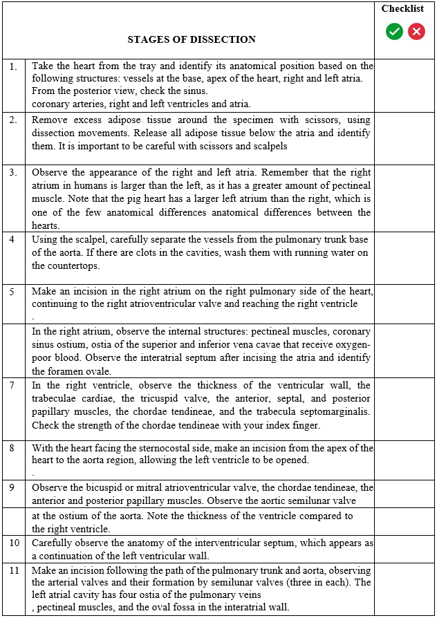
Source: Authors
A presentation was given on the surgical materials belonging to the dissection kits (scissors, anatomical forceps, rat-tooth forceps, scalpel handle No. 4, and scalpel blades), in addition to precautions related to biosafety and the correct use of instruments during the dissection activity (MIZERES; GARDNER, 1988; WEBER, 2001; RODRIGUES, 2010). At each workbench, there was a League monitor to accompany the participants and answer any questions that might arise during the dissection of the heart (Figure 1).
Figure 1 – Anatomy Laboratory and participants during the practical class promoted by LAMAC and monitors from the anatomy course
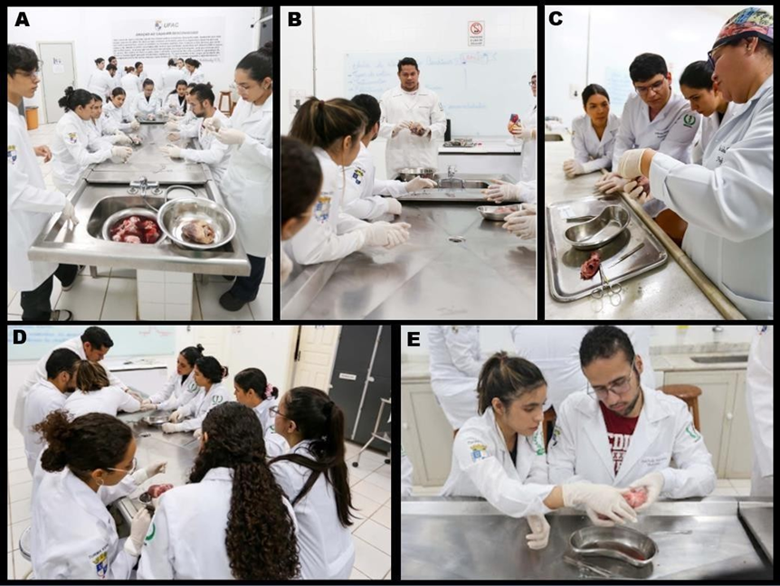
In A and B, we can see the distribution of the groups and explanations about the importance of dissection as an active methodology tool in anatomy. In C, we can see the explanation of the use of the instruments used in the dissection of the pig heart. In C and D, we see the students applying the dissection checklist.
The students followed the dissection steps, beginning with anatomical positioning (Figure 2), thus observing the structures present on the external surface in an anterior and posterior view. After the cardiac anatomical position, the structures were identified in an anterior view: vessels at the base, apex of the heart, right and left atria. In the posterior view, the coronary sinus, ventricles, and right and left atria were verified. In a didactic manner, it was possible to clarify all doubts and thus proceed to the next steps.
Figure 2. Mosaic of the anatomical positioning of the heart
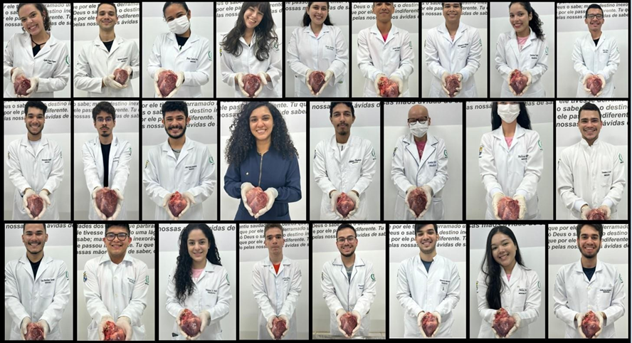
After reading the checklists, the students positioned the heart in the anterior view for recording. It is important to highlight the teamwork of each group in demonstrating the structures.
The students then proceeded to dissect the structures according to the checklist, checking step-by-step the internal structures present in the atrial and ventricular cavities. At the end of the activity, a greater spatial understanding of the heart was observed in relation to the structures located anteriorly and posteriorly, as well as the identification of structures that were not shown in three dimensions (3D) during the demonstration class. During direct observation, active participation, frequent questions, and effective involvement were noted among all groups participating in this event.
Discussion
Heart dissection proved to be an effective teaching strategy, corroborating previous studies that highlight the importance of practical activities for anatomical learning (GONÇALVES et al., 2019; PEREIRA; ROCHA, 2020). Direct contact with the real specimen favors three- dimensional perception and content retention, something limited in two-dimensional (2D) representations or synthetic models that mimic in vivo (MOORE; DALLEY; AGUR, 2024).
In addition to cognitive gains, the activity promoted social-emotional skills, such as cooperation and communication, aligning with the assumptions of active methodologies, which prioritize student participation in the learning process (BERBEL, 2011).
However, it should be noted that the implementation of this practice requires adequate infrastructure, safe handling of biological material, and constant supervision to ensure pedagogical effectiveness and biosafety.
Conclusion
Heart dissection, as an active methodology tool, contributed to meaningful learning, engagement, and the development of practical skills in medical students. The association between theory and practice favored anatomical understanding and clinical reasoning, proving to be a valuable strategy in teaching anatomy.
We suggest expanding similar practical activities to other content areas, with interdisciplinary integration and continuous evaluation of their pedagogical impact. We also suggest expanding to other courses in the health field.
References
BERBEL, Neusi Aparecida Navas. Active methodologies and the promotion of student autonomy. Semina: Social and Human Sciences, v. 32, n. 1, p. 25–40, 2011.
Drake, Richard L.; VOGL, A. Wayne; MITCHEL, Adam W. M.: Gray’s Clinical Anatomy for Students. 4th ed. Rio de Janeiro: Elsevier, 2021.
Frank H. Netter Atlas of Human Anatomy. 8th ed. Rio de Janeiro: Elsevier, 2024.
GONÇALVES, Maria Eduarda et al. The importance of dissection practice in anatomy teaching. Brazilian Journal of Medical Education, v. 43, n. 2, p. 165–172, 2019.
HANSEN, John T. Netter Clinical Anatomy. 4th ed. Rio de Janeiro: Elsevier, 2019.
MOORE, Keith L.; DALLEY, Arthur F.; AGUR, Anne M. R. Clinically Oriented Anatomy. 9th ed. Rio de Janeiro: Guanabara Koogan, 2024.
Paulsen, Friedrich. Sobotta Practical Atlas of Human Anatomy. 3rd ed. Rio de Janeiro: Guanabara Koogan, 2019.
PEREIRA, João Paulo; ROCHA, Carla Cristina. Active methodologies in anatomy teaching: an integrative review. Journal of Health Education, v. 8, n. 2, p. 102–110, 2020.
SILVA, André Luiz et al. The use of dissection as an active methodology in teaching human anatomy. Journal of Health Sciences Education, v. 4, n. 1, p. 45–53, 2021.
1Medical student Federal University of Acre b.rodrigues13pel@gmail.com
2Medical student Assis Gurgacz College University Center
3Medical student Uninorte University Center
4Nursing student Uninorte University Center
5Biomedicine student Unicesumar College
6Medical student Federal University of Acre
7Medical student Federal University of Acre
8Medical student Uninorte University Center
9Medical teacher Federal University of Acre
10education teacher Federal University of Acre
11Coordinator and medical teacher Federal University of Acre