REGISTRO DOI:10.5281/zenodo.12615989
Naiá Taís Magalhães de Carvalho
Abstract
The accuracy of age estimation through cranial measurements is crucial in forensic anthropology and archaeology. This process involves analyzing specific cranial features to determine age, aiding in the identification of skeletal remains and understanding past populations. Technological and methodological advancements have improved the precision and reliability of these estimations. Various studies highlight different approaches: Tobel et al. (2019) showed that combining MRI data from multiple anatomical sites enhances accuracy; Galera, Ubelaker, and Hayek (1998) found methods focusing on endocranial suture closure to be most effective; Lagacé et al. (2019) tested a new histomorphometric method with mixed results; and Serinelli et al. (2015) confirmed the reliability of MRI-based skeletal age estimation in young individuals. These advancements contribute significantly to both historical research and forensic investigations. Other studies were conducted as presented in the work.
Keywords: Age estimation; Forensic anthropology; Cranial measurements; Skeletal identification; Accuracy.
The precision of anthropometric methods for estimating age using the skull is a pivotal area of study in forensic anthropology, archaeology, and related fields. These techniques, which involve the measurement and analysis of specific cranial features, provide essential data for identifying and understanding skeletal remains. Accurate age estimation is crucial for reconstructing demographic profiles of past populations, understanding their health and lifestyle patterns, and solving contemporary forensic cases. Recent advancements in imaging technology, statistical analysis, and morphometric techniques have significantly enhanced the accuracy and reliability of these estimations. By refining these methods, researchers can improve the overall precision of age determination, contributing to more accurate historical reconstructions and supporting justice in forensic contexts.
Reconstructing the biological profile of unknown individuals is incomplete without determining their age. Forensic anthropologists estimate age using skeletal indicators that reflect processes of bone resorption, deposition, and remodeling, which are time-dependent. Estimating age in adults is particularly challenging due to the complexities and individual variations in the aging process and the numerous environmental factors influencing it. As a result, anthropologists typically provide an age range rather than a specific age. It has been observed that the estimated age range for younger individuals is narrower compared to that for older individuals (PRIYA, 2017).
Tobel et al. (2019) conducted a study to assess the performance of age estimation using combined magnetic resonance imaging (MRI) data from all four third molars, the left wrist, and both clavicles, along with anthropometric and sexual maturation data. The study involved 160 female and 138 male healthy Caucasian volunteers aged 14 to 26, who underwent three Tesla MRI from March 2012 to May 2017. Developmental stages were assessed, anthropometric measurements were taken, and self-reported sexual maturation data were collected. A continuation-ratio model was used to estimate age, applying Bayes’ rule for point and interval predictions. The study examined two performance aspects: the accuracy and uncertainty of point predictions, and the ability to distinguish minors from adults (≥18 years). The study concluded that multi-factorial MRI data improve all aspects of age estimation, while anthropometric and sexual maturation data do not provide additional relevant information.
Galera, Ubelaker, and Hayek (1998) examined 963 skeletons from the Terry Collection, consisting of 408 White and 555 Black individuals, to evaluate macroscopic cranial age estimation methods. They applied the techniques of Acsádi and Nemeskéri, Masset, Baker, and Meindl and Lovejoy to each skull. Their findings indicated that the most reliable methods involved assessing endocranial suture closure. Specifically, the methods of Acsádi and Nemeskéri and Masset proved to be the most accurate across all subsamples, including divisions by population, sex, and sex within populations, though their relative accuracy might vary when applied to different populations.
Lagacé et al. (2019) explored histomorphometric methods for estimating age-at-death as an alternative to macroscopic techniques in forensic science. To validate the effectiveness of new methods, they must be tested on independent samples to ensure their accuracy. The primary objective of this study was to evaluate a new age-at-death estimation method introduced by Goliath et al. (2016). The research was conducted on a sample of 29 decalcified femur sections from individuals autopsied at the Institute of Legal Medicine in Montpellier, France. The results showed that the formula proposed by Goliath et al. was not effective in estimating age-at-death in this sample, accurately estimating only four out of 29 individuals. Despite this outcome, the study observed the same age-related histomorphometric trends as Goliath et al., albeit with a lower correlation intensity. The differing bone preparation technique used compared to that of Goliath et al. precludes a definitive conclusion about the accuracy of the method.
In forensic practice, there is a growing need for accurate age estimation methods, especially for young individuals. Serinelli et al. (2015) evaluated Tomei’s MRI method for estimating skeletal age in 12 to 19-year-olds. They analyzed MRI images of the left hand and wrist from 77 males and 74 females. The study found strong inter-rater agreement and high correlation between skeletal and chronological age, with Pearson coefficients of 0.98 for males and 0.97 for females. Bland–Altman analysis showed no significant differences between the two age estimates. The study concluded that MRI-based skeletal age estimation is reliable and strongly correlates with chronological age, making it useful for forensic purposes.
Porto et al. (2020) developed a method that utilizes photo-anthropometric indices, derived from cephalometric landmarks on the frontal faces of the Brazilian population, to construct an artificial neural network classifier for estimating age and sex. The binary classifier achieved notable accuracy, particularly for sex estimation in individuals over 14 years old, with F1 measure accuracy values exceeding 0.85. For age estimation, the classifier attained an F1 measure accuracy of 0.72 with a 5-year age interval. Additionally, for age group estimation, the F1 measures of accuracy were above 0.93 and 0.83 for the 14 and 18-year thresholds, respectively.
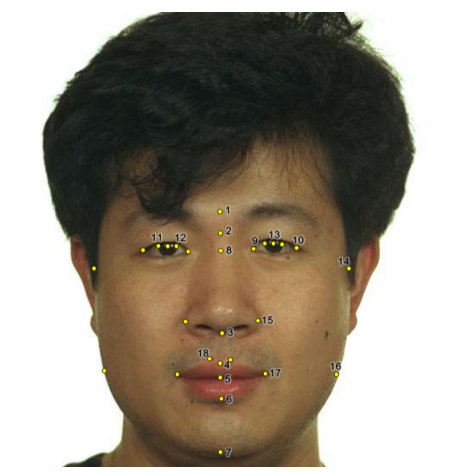
Figure 1: All 28 cephalometric landmarks adopted in Porto et al. (2020).
Lorkiewicz-Muszynska et al. (2015) conducted a study to evaluate the accuracy of anthropometric measurements taken from skeletonized skulls compared to those obtained from their 3D reconstructions via CT scanning. Statistical analysis demonstrated that 3D reconstructions are entirely reliable for any skull measurements. The measurements derived from CT images were accurate and comparable to those from the anthropometric analysis of the skeletonized skulls. There was a strong correlation between the anthropometry of the skeletonized skulls and the CT reconstructions.
In conclusion, the studies reviewed highlight significant advancements in the field of forensic anthropology, particularly in the methods used for age estimation through skeletal analysis. The integration of imaging technology, such as MRI and CT scans, alongside traditional anthropometric techniques, has markedly improved the accuracy and reliability of age determination. These refined methods not only aid in forensic investigations but also enhance our understanding of historical populations and their lifestyles. Despite the challenges, especially in adult age estimation, the continuous development and validation of these techniques underscore their critical role in forensic contexts, contributing to the accurate reconstruction of biological profiles and supporting the pursuit of justice.
References
Galera, V., Ubelaker, D., & Hayek, L. (1998). Comparison of macroscopic cranial methods of age estimation applied to skeletons from the Terry Collection. Journal of forensic sciences, 43 5, 933-9. https://doi.org/10.1520/JFS14337J.
Lagacé, F., Verna, E., Adalian, P., Baccino, E., & Martrille, L. (2019). Testing the accuracy of a new histomorphometric method for age-at-death estimation. Forensic science international, 296, 48-52. https://doi.org/10.1016/j.forsciint.2019.01.020.
Lorkiewicz-Muszynska, D., Kociemba, W., Sroka, A., Kulczyk, T., Żaba, C., Paprzycki, W., & Przystańska, A. (2015). Accuracy of the anthropometric measurements of skeletonized skulls with corresponding measurements of their 3D reconstructions obtained by CT scanning. Anthropologischer Anzeiger; Bericht uber die biologisch-anthropologische Literatur, 72 3, 293-301. https://doi.org/10.1127/anthranz/2015/0481.
Porto, L., Lima, L., Franco, A., Pianto, D., Machado, C., & Vidal, F. (2020). Estimating sex and age from a face: a forensic approach using machine learning based on photo-anthropometric indexes of the Brazilian population. International Journal of Legal Medicine, 134, 2239 – 2259. https://doi.org/10.1007/s00414-020-02346-5.
Priya, E. Methods of skeletal age estimation used by forensic anthropologists in adults: a review. Foresic Research & Criminology International Journal, v. 4, n. 2, 2017.
Serinelli, S., Panebianco, V., Martino, M., Battisti, S., Rodacki, K., Marinelli, E., Zaccagna, F., Semelka, R., & Tomei, E. (2015). Accuracy of MRI skeletal age estimation for subjects 12–19. Potential use for subjects of unknown age. International Journal of Legal Medicine, 129, 609-617. https://doi.org/10.1007/s00414-015-1161-y.
Tobel, J., Fieuws, S., Hillewig, E., Phlypo, I., Wijk, M., Haas, M., Politis, C., Verstraete, K., & Thevissen, P. (2019). Multi-factorial age estimation: A Bayesian approach combining dental and skeletal magnetic resonance imaging. Forensic science international, 306, 110054. https://doi.org/10.1016/j.forsciint.2019.110054.